Bursae and human movement
By R. David Whitby, Contributing Editor | TLT Worldwide January 2023
Friction in our bodies is mitigated by these small fluid-filled sacs.
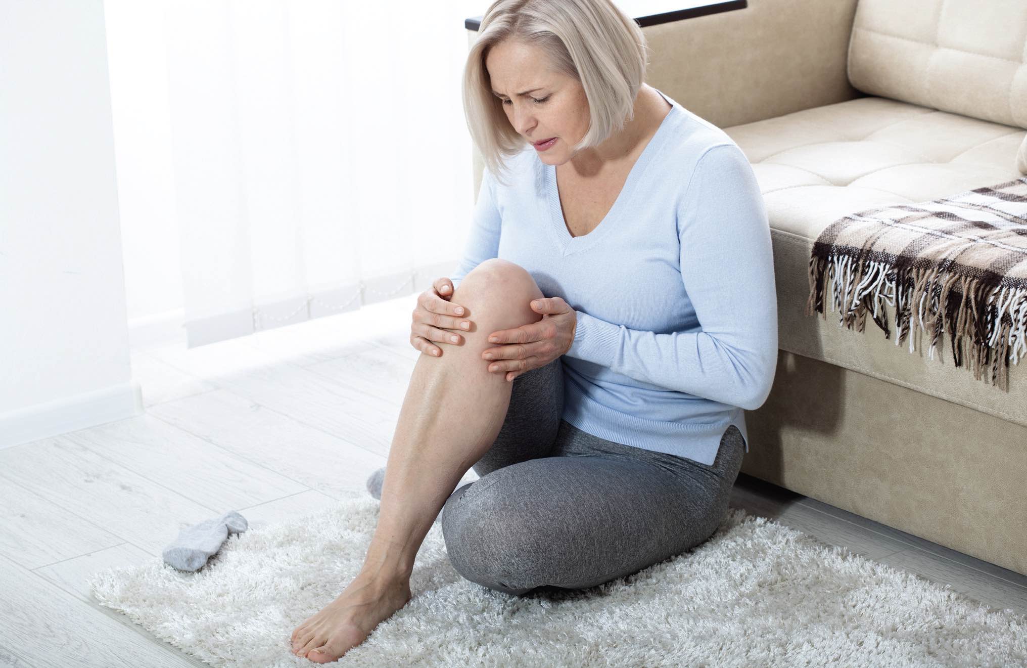
Injury to a bursa may cause pain, limited motion and decreased functional mobility.
Bursae (singular bursa) are small sacs or saclike cavities filled with synovial fluid, in places in the human body in which there could be friction between muscles or tendons and bones or ligaments. They act as a cushion between muscles, ligaments and bones and help to ease the sliding of muscles or tendons over bony or ligamentous surfaces.
There are about 160 bursae in a human body, and they are found throughout. The most important are located in the shoulder, elbow, knee and hip joints. They are very small and thin, with an average diameter in an adult human of around 4 centimeters and a thickness of about 2 millimeters.
Injury to a bursa may cause pain, limited motion and decreased functional mobility. Some injuries may cause a bursa to fill with blood or red or white blood cells. Inflammation of a bursa is called bursitis. A bursa’s membrane is semi-permeable, allowing some materials to flow into and out of the sac. The tissue of the thin membrane secretes the synovial fluid, which is contained within the sac. The viscous synovial fluid, the body’s lubricant, inside the bursae allows the body’s structures to glide over one another with significantly less friction and wear.
A human body has three types of bursae. Synovial bursae are the most common and lie near the synovial membranes of the body’s joints. Submuscular bursae are located between muscles and bony prominences and, in some cases, between neighboring muscles. Adventitious bursae occur only after continued shearing or repeated pressure over a bony prominence. A bunion is an example of an adventitious bursa. Adventitious bursae are not permanent, though they typically form in areas affected by chronic friction, such as in the foot. Subcutaneous bursae lie between the skin and a bony prominence, thereby allowing frictionless motion of skin over bone. An example of this can be found on the back of an elbow. Subcutaneous bursae ordinarily are ill-defined clefts at the junction of subcutaneous tissue and deep fasciae (sheets of fibrous tissue). These bursae acquire a distinct wall only when they become abnormal, when they are sometimes classed as adventitious.
About five bursae surround various areas of a person’s knee joint, providing cushioning. They include the pre-patellar bursa, found overlying the kneecap, the suprapatellar bursa, separating the kneecap from the thigh bone (femoral condyle) and the infrapatellar bursa, found below the kneecap overlying the patellar tendon. A large bursa is located above the bony prominence of each hip joint. This allows a person’s gluteus medius muscles to glide and slide naturally over the bones of both hips. The olecranon bursa lies between the skin and the bony prominence of a person’s elbow. Bursae also are found in people’s feet, to help with cushioning and reduce wear.
Bursitis can occur near joints that perform frequent repetitive motion, such as in the shoulder, elbow or hip. It also can occur in a person’s knee, heel and the base of the big toe.
Treating bursitis usually involves resting the joint, protecting it from further trauma. With proper treatment, the pain subsides within a few weeks, but recurrent flare ups of bursitis are common. People can treat bursitis by trying not to move the joint too much, avoiding activities that put pressure on it, gently holding an ice pack wrapped in a towel on the area for around 10 minutes at a time and repeating this every few waking hours. Paracetamol or ibuprofen painkiller tablets or capsules may help to ease the pain.
If the pain is really bad or does not go away after a few weeks, a doctor may decide to take a sample of fluid from the affected joint using a needle. This will be tested for an infection or medical conditions such as gout. If an infection is found, this can be treated with a course of antibiotics. For other conditions, a steroid injection may be given into the affected joint to reduce the swelling.
If the bursitis is severe or keeps coming back, the inflamed bursa may need to be surgically drained or even removed, although this is rare. If a bursa is surgically excised, it can grow back over a few weeks’ time.
David Whitby is chief executive of Pathmaster Marketing Ltd. in Surrey, England. You can reach him at pathmaster.marketing@yahoo.co.uk.